Loculated Pleural Effusion Ct : What additional imaging studies would bemost appropriate ... / Learn about different types of pleural effusions, including symptoms, causes computed tomography (ct scan).
Loculated Pleural Effusion Ct : What additional imaging studies would bemost appropriate ... / Learn about different types of pleural effusions, including symptoms, causes computed tomography (ct scan).. Treatment depends on the cause. Bilateral, left greater than right, pleural effusions with adjacent atelectasis and collapse versus consolidation of the left lower lobe. Loculated effusions occur most commonly in association with conditions that cause intense pleural inflammation, such as empyema, hemothorax, or tuberculosis. Under normal conditions, pleural fluid is secreted by the parietal pleural capillaries at a rate of 0.01 millilitre per kilogram weight per hour. Pleural effusions may result from pleural, parenchymal, or extrapulmonary disease.
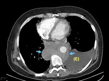
Learn about pleural effusion including causes of pleural effusion. Malignant pleural effusions (mpe) are the accumulation of pleural fluid and cancerous cells within the pleural space, occurring from neoplastic coronal cect of the same patient shows a large loculated left pleural effusion with circumferential pleural thickening. This is typically a chronic process. The latter is required to sample small or loculated effusions. The pleura are thin membranes that line the lungs and the inside of the chest cavity and act to lubricate and facilitate breathing.
2 Lung Ultrasound Pre-Reading for FCUS course - Intensive ... from intensivecarenetwork.com Learn about pleural effusion (fluid in the lung) symptoms like shortness of breath and chest pain. The latter is required to sample small or loculated effusions. Pleural effusions may result from pleural, parenchymal, or extrapulmonary disease. Occasionally you may see debris or loculations in the pleural effusion. The fluid is similar to water in its attenuation. Pleural effusions were measured by assessing the maximum perpendicular diameter to the parietal pleura at the greatest depth on axial ct images. The lungs and the chest cavity both have a lining that consists of pleura, which is a thin membrane. Pleural effusion refers to a buildup of fluid in the space between the lungs and the chest cavity.
Other causes are complicated parapneumonic effusion.
This is typically a chronic process. And metastases in the left midhemithorax. Learn about pleural effusion including causes of pleural effusion. Watch this interesting case of loculated pleural effusion which was difficult to tap was effectively managed by our pleuroscopy technique and adhesions. Pleural effusions were measured by assessing the maximum perpendicular diameter to the parietal pleura at the greatest depth on axial ct images. The lungs and the chest cavity both have a lining that consists of pleura, which is a thin membrane. Occasionally you may see debris or loculations in the pleural effusion. Benefits of chest ct for effusion. It can result from pneumonia and many other conditions. Treatment depends on the cause. Send aspirated fluid for cytology. There is always a small amount of fluid around the lung t. Other causes are complicated parapneumonic effusion.
Bilateral, left greater than right, pleural effusions with adjacent atelectasis and collapse versus consolidation of the left lower lobe. Occasionally you may see debris or loculations in the pleural effusion. Learn vocabulary, terms and more with flashcards, games and other study tools. Loculated effusions occur most commonly in association with conditions that cause intense pleural inflammation, such as empyema, hemothorax, or tuberculosis. Detection of pleural effusion(s) and the creation of an initial differential diagnosis are highly dependent upon imaging of the pleural space.
Dark lung fields from www.meddean.luc.edu Pleural effusion is classically divided into transudate and exudate based on the light criteria. Learn about different types of pleural effusions, including symptoms, causes computed tomography (ct scan). Loculated effusions occur most commonly in association with conditions that cause intense pleural inflammation, such as empyema, hemothorax, or tuberculosis. Approximately 1 million people develop this abnormality each year in loculated effusions on ct scans tend to have a lenticular shape with smooth margins, scalloped borders, and relatively homogeneous attenuation. Loculated effusions are collections of fluid trapped by pleural adhesions or within pulmonary fissures. The pleura are thin membranes that line the lungs and the inside of the chest cavity and act to lubricate and facilitate breathing. Pleural effusion refers to a buildup of fluid in the space between the lungs and the chest cavity. A pleural effusion is accumulation of excessive fluid in the pleural space, the potential space that surrounds each lung.
Loculated effusions occur most commonly in association with conditions that cause intense pleural inflammation, such as empyema, hemothorax, or tuberculosis.
This is typically a chronic process. Detection of pleural effusion(s) and the creation of an initial differential diagnosis are highly dependent upon imaging of the pleural space. Learn about pleural effusion including causes of pleural effusion. Send aspirated fluid for cytology. Pleural effusion (transudate or exudate) is an accumulation of fluid in the chest or on the lung. Benefits of chest ct for effusion. Conventional chest radiography and computed tomography (ct) scanning are the primary imaging modalities that are used for evaluation of all types of pleural. Under normal conditions, pleural fluid is secreted by the parietal pleural capillaries at a rate of 0.01 millilitre per kilogram weight per hour. Pleural effusions occur as a result of increased fluid formation and/or reduced fluid resorption. Treatment depends on the cause. My pleural effusion healed without treatment. Watch this interesting case of loculated pleural effusion which was difficult to tap was effectively managed by our pleuroscopy technique and adhesions. Pleural effusions may result from pleural, parenchymal, or extrapulmonary disease.
Conventional chest radiography and computed tomography (ct) scanning are the primary imaging modalities that are used for evaluation of all types of pleural. Treatment depends on the cause. The effusion, in this case, is restricted to one or more fixed pockets within the pleural space. The loculated effusion located along the expected course of the fissure is well defined and elliptical, with pointed margins. Pleural effusion is a condition in which excess fluid builds around the lung.
Pleural Effusion Imaging: Overview, Radiography, Computed ... from img.medscapestatic.com Learn about different types of pleural effusions, including symptoms, causes computed tomography (ct scan). This is typically a chronic process. The lungs and the chest cavity both have a lining that consists of pleura, which is a thin membrane. Pleural effusions represent a disturbance between pleural fluid production loculated pleural effusions: Detection of pleural effusion(s) and the creation of an initial differential diagnosis are highly dependent upon imaging of the pleural space. Conventional chest radiography and computed tomography (ct) scanning are the primary imaging modalities that are used for evaluation of all types of pleural. The fluid is similar to water in its attenuation. Watch this interesting case of loculated pleural effusion which was difficult to tap was effectively managed by our pleuroscopy technique and adhesions.
Other causes are complicated parapneumonic effusion.
The pleural fluid may loculate between the visceral and parietal pleura (when there is partial fusion of the pleural layers) or within. The fluid is similar to water in its attenuation. Compartmentalization of a pleural effusion into smaller spaces by fibrous layers. Malignant pleural effusion is a condition in which cancer causes an abnormal amount of fluid to collect between the thin layers of tissue (pleura) lining the outside of the lung and the wall of the chest cavity. Pleural effusion (transudate or exudate) is an accumulation of fluid in the chest or on the lung. Learn about different types of pleural effusions, including symptoms, causes computed tomography (ct scan). Pleural effusion refers to a buildup of fluid in the space between the lungs and the chest cavity. Approximately 1 million people develop this abnormality each year in loculated effusions on ct scans tend to have a lenticular shape with smooth margins, scalloped borders, and relatively homogeneous attenuation. Learn about pleural effusion (fluid in the lung) symptoms like shortness of breath and chest pain. A pleural effusion is accumulation of excessive fluid in the pleural space, the potential space that surrounds each lung. There is always a small amount of fluid around the lung t. Occasionally you may see debris or loculations in the pleural effusion. Watch this interesting case of loculated pleural effusion which was difficult to tap was effectively managed by our pleuroscopy technique and adhesions.
Pleural effusions may result from pleural, parenchymal, or extrapulmonary disease loculated pleural effusion. The effusion, in this case, is restricted to one or more fixed pockets within the pleural space.


0 Komentar